CBCT Confirms Chronic Periodontitis, Rules Out Vertical Root Fracture
By: Jason G. Souyias, DDS and Mark K. Setter, DDS, MS
Download PDF Version With Clinical Case Images
Case Overview:
A 48-year-old Caucasian female was referred to our office for a suspected vertical root fracture of tooth #19. The patient was generally healthy, but had previously been diagnosed with generalized moderate chronic periodontitis. The periodontal disease received scaling and root planning therapy in the patient’s general dental office. The patient was placed on 3month periodontal maintenance.
Several years following her periodontal therapy, persistent pocketing was noted on tooth #19 distal, and the patient was referred due to suspicion of a vertical root fracture. Upon examination in our office the patient was found to have generalized 2-3 mm probing depths, with the exception of tooth #19 distal (8 mm) and tooth #22 distal (9 mm with exudate).
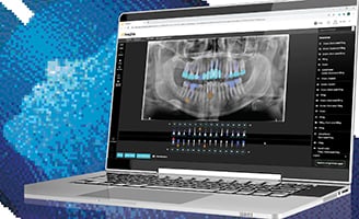
A CBCT scan was ordered to rule out a fracture of the tooth. The scan, conducted with our CS 9300 system, confirmed our suspicion of persisting localized chronic periodontitis as opposed to a vertical root fracture.
Treatment Plan:
A treatment plan was drawn up for guided tissue regeneration around the two sites. The CBCT scan allowed us to know EXACTLY what the defect looked like before surgery. This information helped us to choose more appropriate materials, assign a more accurate fee for the procedure, and predict the success of the outcome more accurately for the patient.
Testimonial:
Periodontal applications obviously require high-resolution images to properly visualize intricate anatomical features. This can be especially helpful for patients whose symptoms are consistent with multiple different types of pathology. In this case, having the CBCT scan at our disposal not only enabled the patient to keep her teeth, but helped us to know what to expect under the gums before we picked up a scalpel. We like that kind of predictability, and many patients prefer a quick, painless CBCT scan over an exploratory procedure.
Further, capturing and analyzing a 3D image of this patient was much faster than – and eliminated the need for – more extensive differential diagnostic methods.


