Iatrogenically Placed Subgingival Elastic Detected by CBCT*
By Robert L. Waugh, DMD, MS, and Assistant Professor of Orthodontics, Medical College of Georgia
Case Overview
This case involves a patient from out of town who was referred to me for evaluation of a possible submerged elastic, which I confirmed through high-resolution 3D CBCT imaging and creative use of the software’s reformatting tools. Interdisciplinary care, facilitated by innovative recorded video communications, was used to accurately diagnose, plan, and implement treatment to correct what could have been a very serious problem.
The patient, 10- year-old Hispanic female, was referred to us by a general dentist on emergency transfer to evaluate a worsening periodontal condition around the upper central incisors [FIGURE 1>. Despite the best efforts, careful periodontal attention was failing as the condition had deteriorated to the degree that it was compromising the patient’s occlusion. Since the patient was in limited orthodontic treatment in another city, she was referred to me for orthodontic evaluation.
Treatment Plan
On examination, the patient shared a history of having placed elastics around teeth numbered 8 and 9— at the supervision of a staff member of her previous orthodontist—to close a relapsing diastema. A consulting periodontist was reluctant to flap the tissue to search for the suspected presence of an elastic without better evidence. A focused-field, high-resolution scan was made and the soft tissue filters were used to reveal the space occupied by the elastic [Figure 2>.
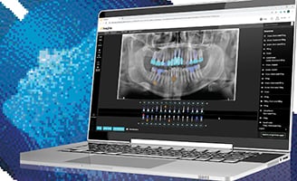
An in-depth video narrative was made of the patient’s case using Camtasia screen capture and CS 3D Imaging Software. This simple setup allows for “on-the-fly” collaboration between treating specialists and related parties—in this case the general dentist, the patient’s parents, the periodontist, and me, as the treating orthodontist. CS 3D Imaging Software imaging made it easy to create the story and more realistically put a face behind the case.
Guided by the video imagery of the cone beam CT manipulation [Figure 3>, the periodontist used a surgical flap to confirm the presence of the elastic, which was removed and a graft placed [Figure 4>. The patient later returned to our care to finalize the occlusal discrepancy.
The periodontist provided a follow-up photograph two months post-surgery and reported they were “very pleased with the outcome” [Figure 5>.
Testimonial
By combining industry-leading, high-resolution CBCT combined with powerful imaging software, a visual narration was used to engage an interdisciplinary team to treat a once-in-a-career problem of iatrogenically-placed elastic with concomitant periodontal destruction. The case ended successfully in what could have otherwise been an unfortunate outcome.
*View a video of this clinical case at www.carestreamdental.com/waughCBCT.


