by Kyle Shank, D.D.S, Shank Center for Dentistry, Indianapolis, Ind.
Challenge: 2D imaging doesn’t always reveal the source of a problem
Solution: CS 9300 CBCT and Panoramic Imaging System
Benefits:
Increased diagnostic capabilities
Expanded treatment/procedures
Enhanced patient education
Greater case acceptance
When patients come to Dr. Shank with a dental problem, they typically don’t have to wait long for a solution. That’s because Dr. Shank prefers a workflow that enables him to present treatment plans to his patients while they are still in the chair. Instead of referring them out for a scan and waiting for a follow-up visit to review it, Dr. Shank has the capability in-house to see all the anatomical detail necessary to make a diagnosis—that is, since he acquired the CS 9300 CBCT and panoramic imaging system.
“The pathology that is readily visible with this system is really quite remarkable,” said Dr. Shank. “I am able to see the full extent of a problem and diagnose accordingly with complete confidence.” Dr. Shank’s patients find the CS 9300’s images to be very impressive, informative—and persuasive. They can see for themselves the status of their clinical situation and quickly comprehend Dr. Shank’s treatment plan.
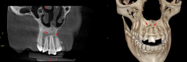
At no time was image clarity more valuable than with a patient who repeatedly came into Dr. Shank’s office complaining of pain in the back of her mouth. “Before we added the CS 9300 to our practice, this patient came in for several routine cleanings complaining of non-specific pain in the posterior maxilla. During each visit, we would tap around and find multiple tender spots, but we could never isolate anything—and neither 2D intraoral nor 2D extraoral images were assisting in the diagnosis. With so many uncomfortable areas all in the posterior maxilla, we kept writing it off as a sinus issues.”
But as soon as the CS 9300 system was up and running in his practice, Dr. Shank brought the patient back in—on New Year’s Eve. “Lo and behold, she had five failing root canals in the posterior maxilla,” said Dr. Shank, “and none of them had shown up with 2D imaging.”
Obviously the patient wasn’t thrilled about the need for so many endodontic re-treatments, but she was delighted to get the problem resolved and is now pain free. This diagnosis is consistent with Dr. Shank’s findings among several other patients. “After using CBCT for over a year now, I am able to clearly see so many things I could not see on my 2D images, like missed canals in endodontic-treated teeth, unpredictable and hard-to-see root-canal anatomy and difficult-to-identify pathology,” said Dr. Shank. “With the ability to see in 3D, I can identify anatomic abnormalities for implant planning. In the past, when 2D results revealed nothing and our clinical exam was less than conclusive, we used to tell people that everything was okay or we would wait for symptoms to change. Now we take a CBCT scan and can give our patients a definitive diagnosis on the spot.”
Increased Diagnostic Capabilities
With four selectable fields of view, the CS 9300 covers a broad range of clinical applications. Dr. Shank can capture the area of interest that he needs to confidently diagnose even the most difficult cases. “We had a patient who was thrown from a motorcycle, and we were able to identify a fractured maxilla,” said Dr. Shank. “Our 2D images didn’t show the full extent of the fracture, but with 3D imaging, we were able to more accurately diagnose the condition and go in with a much more definitive game plan for how to treat this patient.”
Expanded Treatment/Procedures
The benefits don’t stop with diagnoses, however. The new system eliminates guesswork when it comes to treatment planning as well. “No more see-what-we-get-when-we-get-in-there treatment. I know exactly what I’m going to see and how I’m going to treat it ahead of time,” said Dr. Shank. “Not only can I diagnose an endodontic issue, I can actually do more of the endodontic treatment myself because I am able to see canal anatomy in much greater detail. Before I had the CS 9300, I didn’t attempt certain cases due to a lack of detailed information, which made it difficult to see the canal anatomy. Now with more information in three dimensions, I know that I can do some of those cases.”
On the other hand, Dr. Shank can accurately determine which cases not to take on. “You might have a premolar that looks like it has a single canal running right through the middle,” said Dr. Shank. “Prior to CBCT, I would’ve thought, ‘Sure, I can do that one.’ But I’ve seen cases where CBCT revealed that 3 mm from the apex, the canal splits off in three different directions into a C-shaped fin. These are the most challenging cases even for an endodontist with a microscope.”
Enhanced Patient Education and Greater Case Acceptance
Since he acquired the CS 9300, the number of implant surgeries Dr. Shank performs has increased dramatically. He chalks this development up to one thing: confidence. “I am more confident going into the surgery because I’ve already seen the bone three dimensionally and planned the case out ahead of time, and my patients are more confident because of what they’re seeing” he said.
This increase isn’t due to a greater number of extraction diagnoses. Rather, Dr. Shank’s patients are more willing to say ‘yes’ to treatment—in-house. They recognize the planning that Dr. Shank puts into each implant procedure from start to finish, and they feel comfortable that he has the technology to back him up. “They know that I’m going to make their surgery as minimally invasive as possible,” said Dr. Shank. “I can predict with confidence whether or not a bone graft is necessary, and I can show them the surgical guide that I’ve printed on our 3D printer to help ensure an excellent result. Spelling out every detail and showing them the amount of care that goes into their case goes a long way with patients.”
Contrast this approach with the one Dr. Shank had to use prior to installing the CS 9300 in his practice. “Before 3D imaging, I would essentially present a worst-case scenario treatment plan with a lot of variables,” said Dr. Shank. “We would tell the patient, ‘well, we are going to aim for not needing a bone graft, but it’s possible we might.’”
With the CS 9300, however, Dr. Shank can speak more decisively. “Now I am able to tell a patient definitively whether or not a graft or membrane is needed and give them an exact cost for their case. This exudes a lot more confidence in our ability to take care of them comfortably and to be able to predictably give them an outstanding result.”
When asked what his patients think of the new system, Dr. Shank said, “They are very impressed by the technology we have incorporated into our office. It sets us apart from other practices in the area and demonstrates our desire to provide the highest quality of care possible. When we began using 3D technology, our case acceptance immediately went up, and that speaks volumes about how our patients perceive this technology in our office. Purchasing the CS 9300 system has been the single best technology purchase I have made since I’ve been in practice, from both a medico-legal and profitability standpoint.”
© 2024 Carestream Dental LLC. All Rights Reserved

