By Matt Hendrickson, U.S. Orthodontic Director
3D images are an important tool in today’s orthodontic practice. However, capturing cephalometric images is critical for evaluating treatment in orthodontics. To ensure that you are getting the most reliable radiographs possible—and to streamline your practice workflow—it’s important to choose the right imaging system for your office. While there are a number of units on the market that can reconstruct a cephalometric image from a cone beam computed tomography (CBCT) scan, these can take a toll on your workflow as well as introduce legal complications into your practice. Wise practitioners are implementing CBCT systems that give them the flexibility of both 2D and 3D imaging.
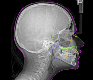 Why Do You Still Need a 2D Cephalometric Unit?
There is no doubt that CBCT technology is growing in popularity in the orthodontic market. While these 3D machines can be used in a number of clinical applications, that shouldn’t discount the place that 2D cephalometric images have in the practice.
“I routinely utilize 3D imaging technology in my practice in the appropriate applications,” said Dr. David M. Sarver, of Sarver Orthodontics in Birmingham, Ala. “However, I feel traditional 2D cephalometric imaging is still the best way to measure growth and treatment response through serial cephalometric superimpositions—the main purpose of cephalometry. I believe it is important for the modern practice to combine both high-quality 2D and 3D imaging in order to maximize treatment quality and practice efficiency.”
The key benefits of using true 2D cephalometric imaging as opposed to cephalometric images reconstructed by a 3D unit include:
Elimination of motion artifacts through one-shot acquisition
Improved workflow
Ability to evaluate treatment response of patients who started treatment with a 2D ceph
Decreased legal liability
Not All 2D Cephalometric Imaging Systems Are Created Equal
Before going into the benefits of 2D cephalometric imaging, it’s important to differentiate between the different systems. Most digital 2D cephalometric units work like a copy machine, meaning they scan from one side of the head to the other over a period of up to 16 seconds. This is actually a step backward from film-based cephalometry, which captured the entire image with one pulse of radiation. This prolonged scan time drastically increases the likelihood of motion artifacts in the 2D cephalometric image. It is critical that clinicians understand this setback and look carefully for systems that offer a “one-shot” ceph. With one-shot cephalometric, one pulse of radiation is generated and the entire image is captured simultaneously. This technology provides a number of benefits, including minimized distortion, improved patient comfort, optimized image quality and reduced exposure time.
Please check back next week, when I will go into more details regarding the advantages of utilizing a 2D cephalometric imaging system within your orthodontic practice.
| Our Family of Brands | ||
 |
 |
|
© 2024 Carestream Dental LLC. All Rights Reserved