CBCT at Its Best: The Perks
Dr. Kunal Shah is the Principal of a new practice in Hendon, London – LeoDental. With a state-of-the-art CBCT installed, the practice is receiving referrals for implant planning cases. As part of a three-part series, Kunal begins by considering the treatment pathway for implant treatment and how CBCT imaging improves the process for a more predictable outcome.
As implant dentistry continues to increase in popularity among the profession and patients, it’s important to establish a protocol for consistently safe and effective treatment. The quality and type of imaging used during the assessment and planning phases have a huge influence on this. In particular, the cutting-edge CBCT scanners now available offer unprecedented visualization of each patient’s anatomy for precise planning and predictable outcomes.
For dentists new to dental implantology, the standard treatment pathway is as follows:
- Patient assessment – One of the most important aspects of treatment, the initial systematic assessment should include everything from review of the patient’s medical history to X-rays. It’s important to evaluate their standard of oral health, home routine, periodontal condition and risk of caries in order to establish whether they are suitable for implant therapy. With peri-implant diseases a big concern in dentistry at the moment, any existing periodontal issues should be treated before implants are even considered.
- Identify all treatment options – Implants should be a listed option when it comes to treatment of failing or missing teeth, but it’s important that patients are aware of all the possibilities. Depending on the clinical situation, this might include bridges, partial or complete dentures, as opposed to implants.
- Treatment planning – Historically, this stage would involve periapical radiographs with a 5mm ball bearing used as a reference point once calibrated. However, with the advancement of technology, a CBCT scan can do everything the CT did and much more. Using the scans, implants can be planned and placed virtually to ascertain positioning and to provide the patient with a visual aid for properly informed consent. An impression should then be taken and a surgical stent fabricated.
- Surgery – Implants are placed, in most cases with a healing abutment. In cases that require bone augmentation, it may sometimes be advisable to place a cover screw, suture effectively and allow suitable time for integration and healing.
- Restoration – Usually about 3-4 months later, following successful osseointegration and sufficient soft tissue healing, impressions can be taken for the final crowns and fitted. With a body of evidence to suggest potential problems with cement, I personally prefer screw-retained restorations to minimise the risks.
- Maintenance – As far as the patient is concerned, this is the most important aspect of treatment – only with their continued compliance to good oral health routines can the best, long-lasting results be achieved.
Benefits of CBCT
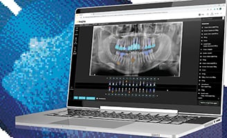
CBCT images show much more than the traditional CT scans, allowing for greater accuracy and predictability during treatment. Both will produce images that identify where the sinus is in the maxilla and where the nerves are located in the mandible. However, the CBCT will also show the bone density, its mesial-distal width, and adjacent space, as well as the buccal-lingual space. This three-dimensional view allows the clinician to determine the suitability of implant treatment, as well as the most appropriate implant type and position. Low bone volume could indicate the need for bone grafting, even at an early stage in the process. I use the CS 8100 3D extraoral imaging system from Carestream Dental, which produces images of outstanding clarity for superior diagnostics every time.
This thorough and more precise planning serves to reduce the risks associated with implant surgery. As such, the actual surgical procedure is quicker and safer than it was using previous planning techniques. The speed of surgery, in particular, is beneficial as it is likely to cause less trauma and therefore promote quicker healing of the soft tissue.
Of course, not every practice has the luxury of a CBCT scanner – the significant investment is not always plausible for practices with low demand for implants or for clinicians who are just starting out in the field. Referral to a center with the latest technology, either just for the CBCT scan or for the whole implant treatment, is a great solution.
Seamless workflow
Another aspect to further improve the workflow and ensure surgery proceeds as smoothly as possible is teamwork. It is crucial that the dental surgeon and dental nurse communicate constantly throughout the procedure to ensure optimal efficiency. It is also important that both professionals are comfortable enough to question the other – whether asking the questions or providing an explanation, the extra thought will help both understand the procedure better. This also means that no one feels unable to point out a different way of doing things or a possible error, further making sure nothing is the missed and the patient receives the very best standard of care and treatment.
With the fundamentals outlined, it’s time to apply this to practice. In part 2, I will demonstrate a specific case and demonstrate how all this can be fully utilized.


