CBCT in the Periodontic Practice

The potential advantages of cone beam computed tomography (CBCT) to periodontology are fairly well known. Implementing a CBCT system into your practice can: streamline the workflow; enhance diagnostic techniques and treatment planning; hone surgical anatomic expertise; and improve outcomes in specific implant and periodontal cases. But because CBCT is a relatively new technology, there’s little evidence-based everyday guidance for the periodontist to rely on.
In the absence of this information—and because CBCT technology is so promising—the American Academy of Periodontology (AAP) used a model of scientific inquiry called best evidence consensus (BEC). BEC is based on the best available current published evidence, as well as expert opinion, and serves as a guide for reasonable uses for CBCT in selected clinical scenarios. In this blog, I would like to highlight several key points from my paper in the Journal of Periodontology, Commentary: Cone-Beam Computed Tomography: An Essential Technology for Management of Complex Periodontal and Implant Cases, regarding the use of CBCT in for three different clinical situations.
Question: Should CBCT imaging replace 2D radiographic analysis when it comes to implants for:
- Assessment of diagnosis and treatment outcome?
- Implant treatment planning?
- Anatomic characterization?
Answer: You’ll find many publications supporting CBCT in all three. Bottom line: CBCT should be used as an adjunct to 2D radiology when the specific benefits to the patient outweigh the risks.
Question: Is CBCT imaging useful in determining risk to periodontal structures in patients requiring tooth movement?
Answer: While there is limited evidence to support CBCT as a routine part of periodontal-orthodontic therapy, experts agree on several scenarios where CBCT should be considered. Patients with:
- A thin dentoalveloar phenotype
- Concomitant recession
Other potential examples include preventative soft tissue and bone augmentation indications and adult versus child applications. Further research is needed.
Question: Does CBCT imaging add clinical value in diagnostic assessment and treatment planning for the management of periodontitis?
Answer: At the present time, a 2D full-mouth radiographic series combined with clinical probing remains the gold standard for comprehensively evaluating periodontal structures. However, experts on the BEC panel identified several situations where the addition of CBCT imaging would be useful, including:
- When advanced furcation lesions have been detected and dental implants are being considered as an alternative treatment option.
- When there is a questionable root fracture, root resorption or periodontal-endodontic lesion present that could not be clearly identified by 2D imaging and/or clinical evaluation.
Minimally invasive therapies and evaluation of regenerative procedure outcomes are two areas of potential benefit. Further research is indicated here, too.
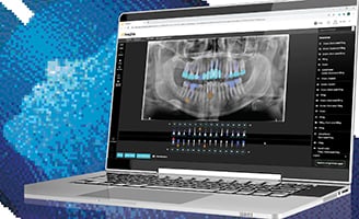
Clearly, CBCT has the potential to improve today’s standard of care. The 3D views CBCT provides can significantly change the course of treatment. New imaging software is further enhancing the use of CBCT, making it a more intuitive and effective tool for treatment planning. In addition, the digital file format of CBCT scans is easily transferable when working with referrals.
Research demonstrating the value of CBCT is ongoing. Ultimately, however, each clinician must assess the benefits and limitations of the technology and the readiness of the practice in determining whether it is appropriate to incorporate it.


