Interpreting Advanced Imaging: It’s Best to Know Nothing
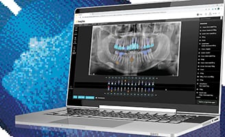
Interpreting advanced imaging, such as CBCT imaging, is tricky. Evidence of just how tricky becomes apparent during lectures I give on the subject. I will show a study to the audience and discuss it for several minutes. When I take it down, I ask them which side was buccal? Which side was palatal? Was it tooth number 14 or number 3?
The audience will start guessing because they really don’t know—even after looking at the tooth for 5-10 minutes. They would never mistake buccal for palatal, or the tooth numbers, with 2D radiography. But with 3D radiography, it happens all too often. I’ve been evaluating 3D imaging every day in my endodontic practice for eight years now, and I can still make this kind of mistake. The issue is this: we don't have that same set of skills with 3D radiography that we do with 2D radiography, but we think we do.
In medical radiology, however, they’re well aware of the complexities that go along with interpreting imaging. That’s why medical radiology is a four-year specialty after medical school—with sub-specialties after that. You don't want a mammographer evaluating the CT of your head or a thoracic radiologist reading your mammogram.
The need for specialization is clear: interpreting 3D imaging calls for a well-trained eye. That’s why we have to be very careful when we interpret with CBCT. For this reason, I’ve developed a strategy for interpreting 3D imaging based on what has been learning in medical radiology.
It’s important to note that I don’t automatically order an imaging on every patient. Whenever possible, my staff provides me with the minimum information necessary to determine if an advanced imaging study should be prescribed. This very counter-intuitive finding is captured in the title of a 2002 paper in Radiology from noted radiologist Dr. Thorn Griscom: “A Suggestion: Look at the Images First, Before you Read the History.”
Whenever possible, the preferred method involves doing two reads—first, without looking at the projection radiograph, doing a clinical exam or talking to the patient first about their symptoms or getting the history. My goal is to not have any preconceived notions about what the findings may be, let alone the diagnosis. Of course, with CBCT—especially with the focused field—if it's an upper left side, I have a pretty good idea of where the problem is. But that's all I really want to know.
I evaluate the study through that lens. I then get the history, look at the projection radiograph, review all the clinical information and perform the clinical exam. After that, I go back and look at the CBCT study again. This approach is very counterintuitive and not widely appreciated. Current recommendations for approaching are as follows: conduct a thorough clinical exam and radiographic exam before prescribing imaging. In my opinion, that’s backwards, and not based on what has been learned about the interpretive process through careful research in medical radiology.
Much of this stems from the division of labor in medicine; the ordering clinician, the interpreting clinician and the intervening clinician are three different people. But for endodontists, we're all three of those clinicians wrapped up in one.
It's not clear to me what the absolute 100-percent best approach is, as we haven’t studied it very well. However, our preliminary work at St. Louis University confirms what has been found in medicine: experienced clinicians missed clear and compelling findings with history that were found without history. As Griscom writes: “The history helps. Interpreting images while unaware of the patient’s problem is foolish. If you aren’t sure what the question is, it’s tough to give the right answer. Sometimes, however, the history misleads.” Endodontists need to keep these issues in mind; that we are all three of those players, and our interpretation of the radiographic imaging can be profoundly biased by what we know before we do the evaluation—both positively and negatively.


