With the evolution of technology, integrating digital workflow embodies the phrase “work smarter, not harder.” Making the laboratory workflow more digital allows clinicians to save time and money without sacrificing a high-quality outcome. When performing a restoration or providing a prosthesis, it is my goal to provide patients with a long-lasting and ideal restoration. With a digital workflow, the delivery of restoration is not only quicker but also decreases the chances of corrections and remakes.
In a recent case, I worked with a 78-year old female patient who had decay and fracture under a PFM Crown. The crown had recently come loose, and the remaining tooth was non-restorable. The treatment plan was to surgically remove tooth # 18, degranulate the site, place a bone graft and cover with a non-resorbable membrane. After implant placement and osseointegration, a digital impression was taken and a natural tooth zirconia crown and an implant bridge were designed and fabricated.
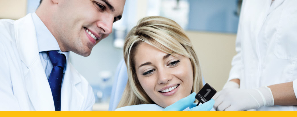
To begin the procedure, the temporary crown was removed from tooth # 18 and cleaned. Next, the BioHorizon healing heads were removed from implant #s 19 and 21 and the gingival collars showed nice petechiae and appeared healthy. New Image Dental lab fabricated abutment seating jigs were utilized to align and seat the custom abutments onto sites # 19 and 21. The abutment screws were then hand-tightened, and the seating jig was removed. An intraoral sensor was used to capture a digital bitewing image to verify proper alignment and seating of the abutments. After this was confirmed, the titanium screwdriver was then utilized to further tighten the abutments to seat.
The 3-unit zirconia implant bridge with retainers (implant abutment #s 19 and 21) and pontic (# 20) were then seated and the margination verified; this was followed by seating and verifying the natural tooth # 18 zirconia crown margination. The restorations had nice interproximal contacts and only needed a very slight occlusal adjustment on the opposing teeth #s 12 and 13.
In the end, the patient was very pleased with the results. The crown and bridge were then removed, and the abutment screw holes were filled with plumbers tape before cementation with Kerr Temp Bond cement. The zirconia crown for tooth # 18 was cemented with Kerr Maxcem Elite resin cement.
Using this case as an example, various tools were used to simplify the workflow and deliver predictable restorative outcomes. Modern intraoral imaging allowed digital impressions to be sent to the lab within minutes, cutting down turnaround times and reducing the number of fabrication steps. When compared with conventional dentistry, a digital workflow allows us to complete a case in fewer steps while enhancing the patient’s comfort and satisfaction.
If you are interested in learning more about utilizing a digital workflow to plan and fabricate prosthetics, I have been invited to speak at the 9th Annual High Altitude Comprehensive Implant Symposium. The symposium outlines implant treatment planning, site preparation, surgical procedures and techniques including sinus elevation, sandwich bone augmentation, full arch immediate reconstruction, esthetic zone implants, Digital Technology & Implant Surgery, and much more!
© 2024 Carestream Dental LLC. All Rights Reserved

