Diagnostic Imaging Made Clear
By
The Digital Stream
| April 13, 2021
The diagnostic process is arguably the most important part of dental treatment. After all, unless a problem is correctly identified it’s impossible to provide a solution that not only solves the issue effectively but that also meets patient expectations.
However, taking an accurate diagnostic image is not always straightforward. There are a number of factors that can impact image quality, and professionals should be aware of these in order to reduce the number of retakes needed.
Why retakes are bad news
There are many reasons why retaking diagnostic images is not ideal. First, we must consider that asking the patient to repeat the imaging process can have a negative impact on their overall experience. A retake can come across as unprofessional, and this could impact the level of trust they have in your services, making them unsure of your capabilities.
Plus, we must consider the dosage of radiographs. It’s always a good idea to limit radiation exposure as much as possible.
There’s also the cost and time consideration that retakes entail. Dental appointments can often be time sensitive, so using this time to take another diagnostic image instead of moving forward with the case is not time well spent. Depending on what type of diagnostic image you’re taking, material costs can come into play, too, leaving you out-of-pocket if you need to take more images than necessary.
However, it is worth mentioning that retakes are necessary in some situations, especially if you are unsure of what the first image is telling you. It’s certainly better to rule out any uncertainty, because if you miss something you may be liable in the eyes of the law. As such, you want to do all in your power to ensure that images offer accuracy and utmost clarity the first time.
Mistakes caused by inaccurate images
Mistakes caused by inaccurate images are common. A study that evaluated how often radiographs are misinterpreted found that dental professionals misdiagnosed the depth of caries up to 40 percent of the time and—in 20 percent of cases—misdiagnosed sound teeth as carious.[i]
These errors in judgement are likely caused by images not being clear enough to effectively show the dentition and any abnormalities. But how can you ensure that your diagnostic images are more accurate?
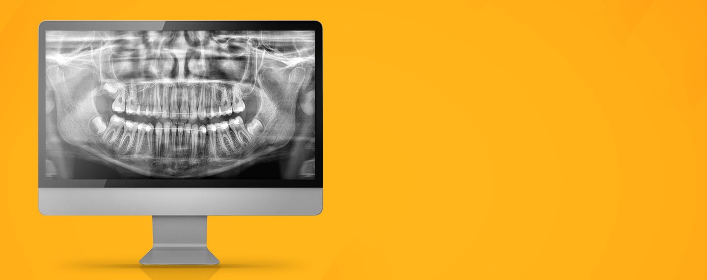
Patient positioning
Part of achieving a good diagnostic image is ensuring that patient positioning is perfect. This is easier said than done, especially if you are treating a patient who is nervous and cannot stay still.
Positioning isn’t just about the body, but also the position of the tongue and other aspects. Positioning errors are incredibly common; in fact, one study assessed approximately 1,800 panoramic radiographs and found that patient positioning errors occurred in a staggering 89 percent of the sample. This meant that almost 25 percent of the images were unacceptable for use in diagnosis and a retake had to be performed.[ii]
In order to limit patient positioning errors, you need to be very clear when telling patients how best to hold their tongue and body in order to get the clearest image. Explain why it is so important and what impact it will have on the efficiency of their treatment. Furthermore, you should look at technology that assists patient positioning—there are some CBCT systems on the market that have this as an in-built feature.
Metal artifacts
Another common enemy to image quality is the appearance of metal artifacts. These are caused by metal in the mouth hardening the X-ray beams used during image acquisition, often resulting in portions of the image being unreadable.
Fortunately, there is technology that has integrated features to combat metal artifacts. For example, the CS 8200 3D from Carestream Dental has a built-in CS MAR module that has been specifically designed to help reduce metal artifacts on images, leading to more precise diagnoses. The system also facilitates laser-free, face-to-face patient positioning, helping to ensure that no mistakes occur during acquisition.
A clearer insight
Diagnostic imaging is essential in order to treat patients effectively. As such, it makes sense to invest in systems that can help overcome common challenges to image clarity, especially if you want to ensure that your patients are receiving the best care possible.
[ii] Dhillon, M. et al. Positioning Errors and Quality Assessment in Panoramic Radiography. Link: https://www.ncbi.nlm.nih.gov/pmc/articles/PMC3534173/ [Last accessed March 21].