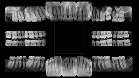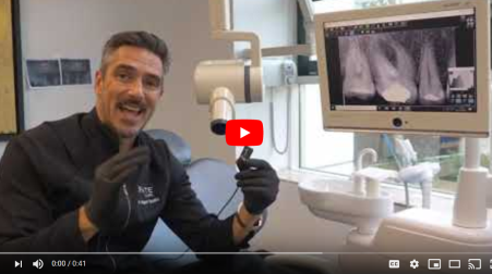
Learn more about customizable technology with the RVG 6200
Meet the RVG 6200—the digital intraoral sensor that adapts to you. From a simplified workflow to user-defined image processing tools, the RVG 6200 is designed to work for you (not the other way around).And, with its straightforward installation process and integration with most imaging and dental practice management software, the RVG 6200 fits easily into any practice.
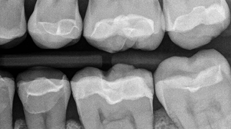
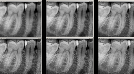
Clinicians can optimize image contrast according to their diagnostic needs or visual preferences with the CS Adapt module—the new feature that lets practitioners establish their own default settings for personalized image processing. Users can choose from either 40 pre-set image enhancement filters or define up to four their own favorite settings, resulting in a customized comfort zone for every appointment.

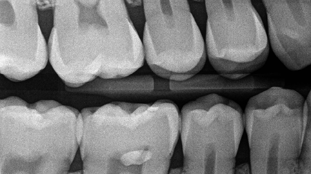
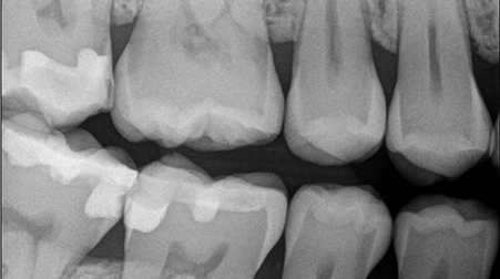
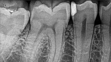
Offering a number of user-defined image processing tools, the RVG 6200 allows you to customize images according to your own specifications for enhanced diagnosis and ease of use.
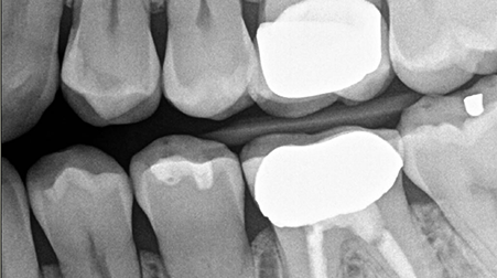
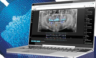
Three anatomical image enhancement modes can be applied to acquired images, including endodontic, periodontic, and dentin-enamel junction, and a user-friendly sharpness filter with dynamic slider bar makes it easy to see contrast changes in real time. Using sharpness ranging from 0 to 6, you can choose the image contrast you prefer.

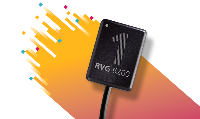
The RVG 6200 sensor features an ergonomically optimized rear-entry cable that reduces bulk at the cable point of entry, allowing for easier placement and positioning of the sensor, improving image acquisition. Additionally, the newly designed reinforced cable is 20% thinner than previous RVG sensors to facilitate better sensor placement in the patient’s mouth. RVG 6200 sensors include a set of RVG 6200 paddle positioning devices so the sensor can be immediately implemented into the practice.

Offering a number of user-defined image processing tools, the RVG 6200 allows you to customize images according to your own specifications for enhanced diagnosis and ease of use.
Three anatomical image enhancement modes can be applied to acquired images, including endodontic, periodontic, and dentin-enamel junction, and a user-friendly sharpness filter with dynamic slider bar makes it easy to see contrast changes in real time. Using sharpness ranging from 0 to 6, you can choose the image contrast you prefer.


