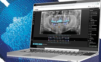
Clinical Possibilities of CBCT
By Dr. Alan Slootsky
Before implementing a cone beam computed tomography (CBCT) unit into my office, there were a few obstacles I had to overcome—but when I did, I found there were a number of things I could clinically accomplish in my office without sending patients out for a scan.
Implant Planning and Placement
CBCT is the standard of care when it comes to planning and placing implants. If an osteotome is needed, a scan allows us to prove that there was no infection pre-op. We can also see the buccal/labial plate thickness and better understand the timeframe if the case is an immediate placement or a graft and wait. In addition, it allows for same-day planning for patients who are visiting the area for a short period and time is an issue.
Of course, CBCT leads to competence—and competence leads to getting the case accepted. Because I can show the patient exactly what is planned on the 3D imaging software and quote the fee upfront, they are able to make well-informed decisions regarding their treatment.
Endodontic Infections
There is no other way to find hidden infections from teeth that either had endo or need endo—especially on the upper teeth, where the zygomatic arch can make taking a periapical image difficult.
With CBCT, we can see sinus infections and any possible relationship to the tooth. In addition, we can definitively see failing endodontic work and identify the problem area. No one wants to take out a tooth that a patient has invested time and money into and—if the clinical suspects a problem with failing endo—he or she wants to be as sure as possible before recommending and proceeding with treatment, from both an ethical and legal viewpoint.
CBCT helps greatly in this area, and when we can find an answer, the patient is so relieved to find that there was an infection.
Oral Surgery
CBCT is critical in risk assessment for third molars. In most cases, we are performing more third molar procedures, we can see the nerve in relationship to the root apex. Having 3D images also allows us to identify cysts and other anatomical structures.
TMJ Cases
Hidden infections can extrude from the tooth and alter the bite, having an impact on the temporomandibular joint. Capturing a scan in TMJ cases aids us in making the right diagnosis and treatment plan.
Resorption
CBCT not only make root resorption easier to diagnose, but deciding on the treatment is easier and the prognosis is improved. Not only that, it is easier to show the problem to the patient with 3D than a flat 2D image.
All in all, CBCT has been a wise investment for my practice by improving diagnostic capabilities, treatment planning and patient communication. How has 3D imaging benefitted you from a clinical perspective?
About Dr. Alan Slootsky D.M.D., M.A.G.D., F.A.C.D.
Born and raised in Bayonne, New Jersey, Dr. Alan Slootsky attended Rutgers University and graduated from the College of Medicine and Dentistry (now called Rutgers School of Dental Medicine). Dr. Slootsky obtained a Fellowship from the Academy of General Dentistry and a Mastership of the Academy of General Dentistry.
Today, Dr. Slootsky is the owner of a successful restorative and cosmetic dentistry practice in Pompano Beach, Fla., as well as a co-chair of the advanced crown and bridge class at the Atlantic Coast Dental Research Clinic. He is a member of several professional organizations, including the American Dental Association, Florida Dental Association, Broward Dental Association, Southern Academy of Prosthodontics and the Pankey Institute, and is a Fellow of the American College of Dentists. He presently is an adjunct professor at Nova University.


